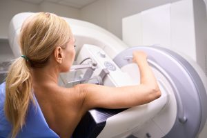
Ultrasound-guided breast biopsies are used by doctors to check for malignant tissue. Your doctor might suggest an ultrasound biopsy if an area in the breast exhibits one or more concerning characteristics. This procedure is usually relatively painless, aside from the pinch and burn of the local anesthetic entering the breast. An ultrasound-guided breast biopsy takes about an hour to complete from start to finish.
What is an Ultrasound-Guided Breast Biopsy?
An ultrasound-guided breast biopsy is a procedure that involves the use of sound waves to locate and accurately sample an abnormality or lump in the breast tissue. The procedure involves the removal of breast tissue samples for examination under the microscope. The tissue samples are acquired through a hollow needle, which makes the procedure much less invasive than a surgical breast biopsy. Once the breast tissue samples are extracted, they are examined by special doctors called pathologists.
Common Uses of the Procedure
Ultrasound-guided breast biopsy procedures have several uses. In medicine, an ultrasound-guided biopsy procedure is carried out when a breast ultrasound reveals an abnormal tissue condition.
When a doctor feels that you may have a lump in your breast or that an area in the breast is displaying concerning characteristics, an ultrasound-guided breast biopsy is typically suggested. It is also utilized to look into odd results on mammograms or other types of breast exams.
What to Expect from the Procedure
Many women report little or no breast discomfort or scarring after the biopsy. However, certain individuals may be more sensitive during the procedure, such as those with thick breast tissue or anomalies along the chest wall or beneath the nipple. In such cases, the patient may notice more discomfort after the biopsy.
You will feel a pinprick from the needle followed by a minor stinging sensation from the local anesthetic when it is used to numb the area. When the doctor enters the biopsy needle and takes tissue samples, you will most likely feel some pressure. This is very normal. Within a few seconds, the region will go numb.
While the doctor does the imaging and biopsy, you must stay completely motionless. The sampling equipment may make clicks or buzzing noises while tissue samples are gathered. These are all very normal.
If you have swelling or bruising after your biopsy, your doctor may recommend that you take over-the-counter pain medicine and apply a cold pack.
How does the procedure work?
Ultrasound is used by doctors to identify changes in the appearance of organs, tissues, and arteries, as well as abnormal structures like tumors.
A transducer delivers sound waves and captures the echoing (returning) waves in an ultrasound test. The transducer transmits short pulses of inaudible, high-frequency sound waves into the body when it is rubbed against the skin.
The sensitive receiver in the transducer captures minute changes in pitch and direction when sound waves bounce off interior organs, fluids, and tissues. These characteristic waves are quickly measured by a computer and shown on a monitor as real-time visuals. Typically, the technologist records one or more frames from the moving visuals as still photos. Short video loops of the photos may also be saved.
The radiologist introduces a biopsy needle through the skin, moves it into the region of the targeted discovery, and extracts tissue samples while using an ultrasound probe to visualize the site of the breast lump, deformity, or aberrant tissue change.
When doing a surgical biopsy, ultrasonography may be utilized to guide a wire straight into the desired discovery to assist the surgeon in locating the region for excision. The clinician can see the biopsy needle or wire as it progresses to the area of the lesion in real-time using continuous ultrasound imaging.
How The Procedure is Performed
Ultrasound-Guided Breast Biopsy is performed while you are laying on your back with your arm lifted over your head. A special, warmed gel is applied to your breast before the biopsy. Then a transducer is put on your breast and carefully pushed back and forth to pinpoint the exact region to biopsy.
The radiologist will clean the region and inject a local anesthetic after it has been detected. A little incision will be made after the region is fully numb. Several cores of tissue will be extracted and submitted to the laboratory for assessment by a pathologist to ensure an appropriate sample is collected. After that, a tissue marker will be applied. You will be informed about the operation and cared for by a nurse.
How Long Does Ultrasound Guided Breast Biopsy Take?
The complete ultrasound-guided core biopsy procedure should take no more than one hour.